European funding for Rubén Fernández Busnadiego
(umg/mbexc) Prof. Dr. Rubén Fernandez Busnádiego from the Institute for Neuropathology at the University Medical Center Göttingen (UMG) and member of the Cluster of Excellence ”Multiscale Bioimaging: from Molecular Machines to Networks of Excitable Cells“ (MBExC) received a Consolidator Grant of the European Research Council (ERC). The ERC supports his research project ”In situ structural basis of human axonopathies“ (“cryoNERVE“) over five years with a total of around 2 million euros. The aim of his research project is to extend the potential of cryo-electron tomography (cryo-ET) to image native nerve tissue at unprecedented resolution. This is necessary to analyze in detail the processes leading to the development of human axonopathies, such as hereditary spastic paraplegia (HSP) and Charcot-Marie-Tooth disease (CMT).
Neurological disorders represent a major health burden and causal treatments are scarce. Neurons face the major challenge of remaining functional throughout our entire lives. Within neurons, axons are particularly vulnerable due to their distance from the neuronal cell body, where most biosynthetic activity takes place. This makes the long axons of motor and sensory neurons (in some cases extending for more than a meter) selectively degenerate in a group of diseases collectively known as axonopathies. In some of these diseases the myelin sheaths, which enable the axons to transmit stimuli in the form of action potentials over otherwise insurmountable distances, are damaged or destroyed.
Major causes of familial axonopathies like HSP and CMT disease are mutations in genes coding for membrane-shaping proteins. Current techniques lack the resolution to ascertain the functions of these proteins and their pathological dysfunction within native nerves. At the same time, the highly specialized architecture of axons and their delicate interplay with glia can only be studied in situ.
PROJECT ”cryoNERVE“
”The basis for the ”cryoNERVE“ project is the overarching hypothesis that membrane morphology crucially determines axonal health and function,“ says Prof. Fernández Busnádiego. ”The further development of methodology for cryo-electron tomography will enable us to image native nerve tissue at unprecedented resolution. This is important to understand how cellular functional units work and are regulated, and how their dysfunctions lead to disease.“ The expert for cryo-electron microscopy has already demonstrated that cryo-ET is a powerful technology to reveal the structural basis of neuronal function and disease in intact cells. However, cryo-ET imaging of tissues remains a major challenge. In the frame of the “cryoNERVE” project, Fernández Busnádiego and his team plan to develop cryo-ET workflows to image and analyze in detail native nerve tissue from mice and flies at unprecedented resolution.
”We will build on these advances to study the function of HSP and CMT proteins within truly physiological environments“, says Prof. Fernández Busnádiego. “Our aim is to answer central research questions: 1) How do these proteins shape axonal organelles and their cross-talk? 2) What is the molecular architecture of myelin sheaths? 3) What are the mechanisms of communication between neurons and glia cells? The alterations introduced by axonopathy-causing mutants will also be analyzed to deepen our molecular understanding of these diseases.
Moreover, Fernández Busnádiego and his team plan to pioneer the first cryo-ET analyses of human tissues preserved into a glass-like state by shock freezing (vitrification). Since peripheral nerve biopsies from axonopathy patients often cannot be diagnosed with existing methods, they will examine patient biopsies at molecular resolution, and capitalize on their findings in flies and mice to reveal hitherto obscured pathological abnormalities.
The project “CryoNERVE” will thus provide a holistic molecular and structural basis of human axonopathies like HSP and CMT, pave the way for high-resolution analy-ses of native human tissues and explore the potential of cryo-ET as a diagnostic tool with nanometric precision.
Prof. Dr. Rubén Fernández Busnádiego is expert in the field of biomedical cryo-electron microscopy that he uses to study the molecular architecture of neurons. After research stays at the Yale University and the Max Planck Institute for Biochemistry in Martinsried, he was appointed as Professor for Structural Cell Biology at the University Medical Center Göttingen in 2019 as part of the MBExC. Within the Cluster of Excellence, he contributes within the with his cross-scale imaging to overcome the boundary between structural and cell biology. His research relies on high-end instrumentation for cryo-electron microscopy/tomography. The costs for the microscopes and the necessary reconstruction of the premises were covered by the German Research Foundation (DFG), the federal government and the state of Lower Saxony.
About the ERC CONSOLIDATOR Grant
With the ERC Consolidator Grant, the European Union supports excellent scientists who are at a career stage where they may still be in the process of consolidating their own independent research team or program. Successful applicants must demonstrate the groundbreaking nature, ambition and feasibility of their scientific proposal. A list with all awardees in this round can be found at: https://erc.europa.eu/sites/default/files/2023-01/erc-2022-cog-results-all-domains.pdf.
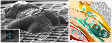
Figure Legend: 3D rendering of the cryo-ET of Drosophila peripheral nerves. Left: Entire nervous system of a fruit fly (Drosophila melanogaster) larva frozen on an EM grid upon dissection. The inset shows a peripheral nerve during thinning by cryo-focused ion beam milling. This process results in thin lamellae of the tissue where high quality cryo-electron tomograms can be recorded. Scale bar: 100µm. Right: 3D rendering of a cryo-electron tomogram of a peripheral nerve. Warm colors represent plasma membranes of different cells: yellow (perineurial glia), orange (sub-perineurial glia), and red (neuronal axon). Organelles are shown in cyan (endoplasmic reticulum), blue (vesicles) and green (mitochondria). Scale bar: 250 nm. Reproduced from https://www.biorxiv.org/content/10.1101/2021.04.14.437159v2.
FORTHER INFORMATIONEN
University Medical Center Göttingen, Georg-August-Universität
Institute for Neuropathology
Prof. Dr. Rubén Fernandez Busnadiego
Justus-von-Liebig-Weg 11, 37075 Göttingen
Phone +49-551 / 39-60745
ruben.fernandezbusnadiego(at)med.uni-goettingen.de
about RG Structural Cell Biology: https://neuropathologie.umg.eu/forschung/arbeitsgruppen-labore/ag-fernandez-busnadiego/
about the MBExC: https://mbexc.de/
Cluster of Excellence Multiscale Bioimaging (MBExC)
Dr. Heike Conrad (Science communication)
Phone +49-551 / 39-61305
heike.conrad(at)med.uni-goettingen.de